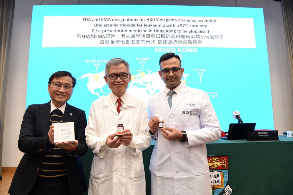
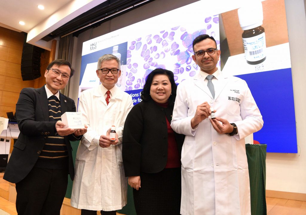
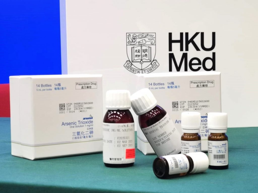
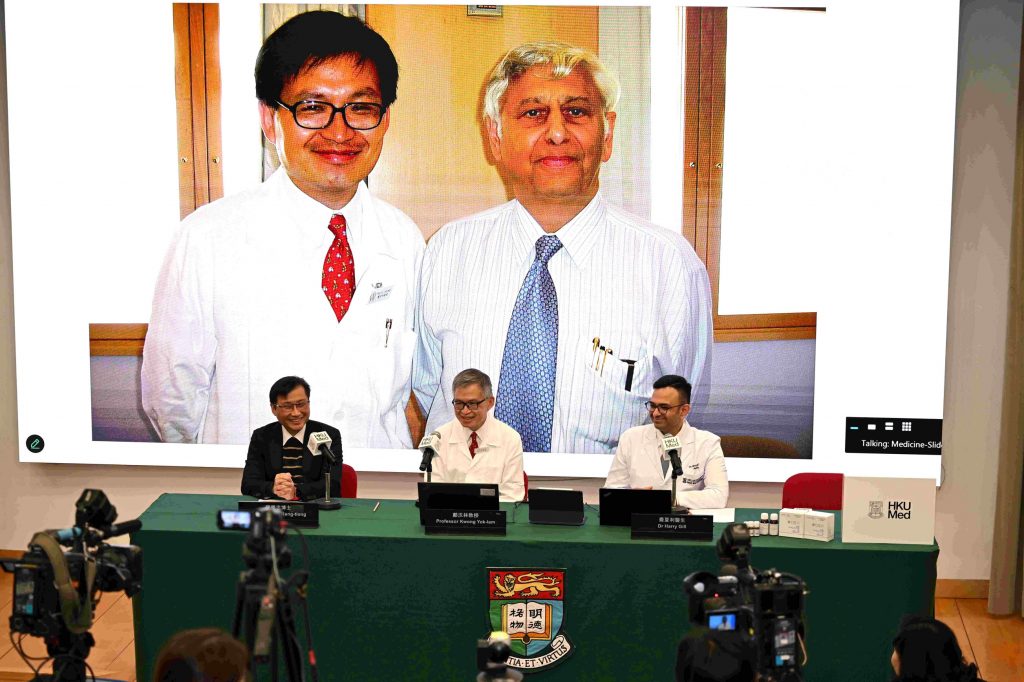
作為將藥用口服砒霜推向國際醫療舞台的第一步,該藥已獲得美國食品及藥物管理局(FDA)及歐洲藥品管理局(EMA)的罕見病藥物資格認定(孤兒藥,orphan drug designation,ODD),亦同時取得美國 FDA新藥臨床研究資格認定(investigational new drug designation,IND)。這是首款由香港研發的抗癌處方藥物,獲得重要的FDA和EMA認證,對進行針對藥用口服砒霜的全球性研究至關重要。
APL 亞洲聯盟
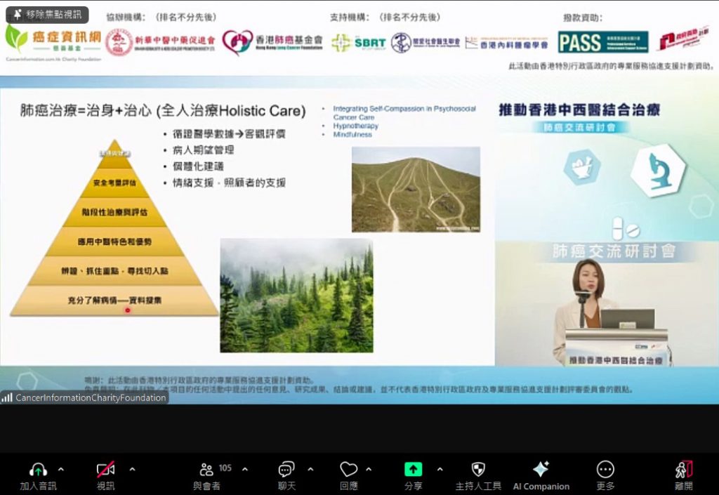
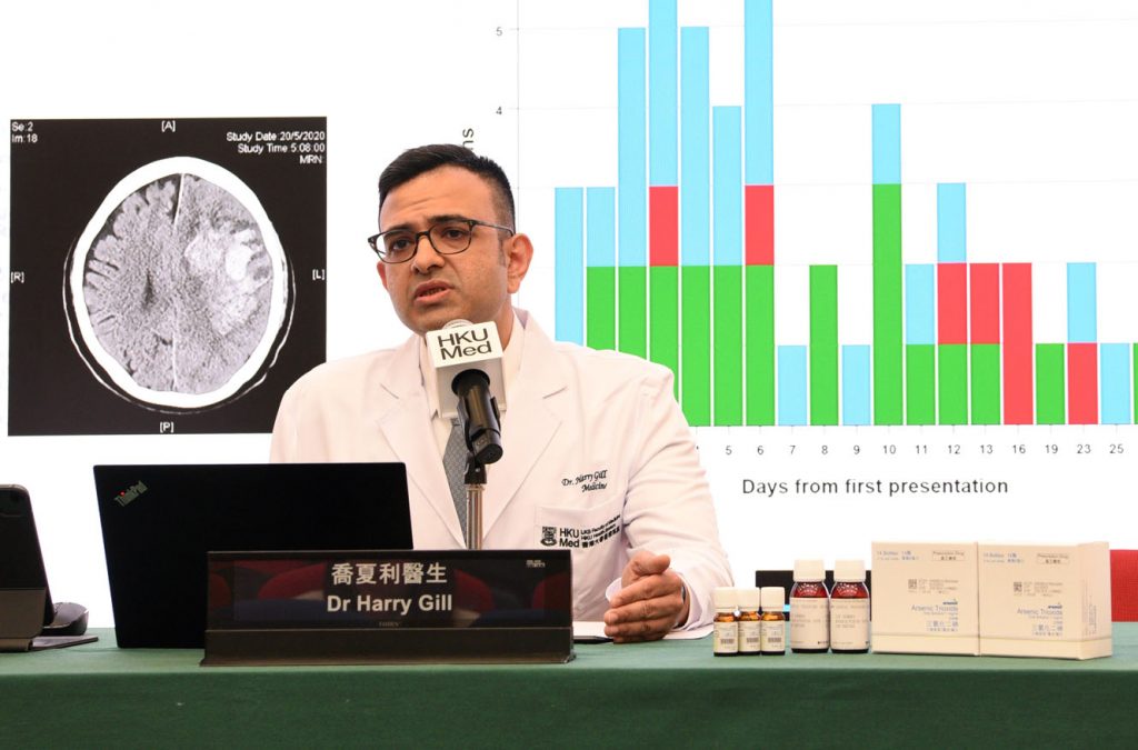
由港大醫學院領導的APL亞洲聯盟 (APL Asian Consortium)由香港、馬來西亞、新加坡和台灣的研究人員組成,專注於APL的研究和治療。在其首個回顧性分析中,發現「AAA」較化療等傳統治療方案,更具優越性。APL 亞洲聯盟是目前在這些地區推廣藥用口服砒霜應用的重要平台,其在亞洲聯合進行的「AAA」研究項目獲港府創新及科技基金支持。
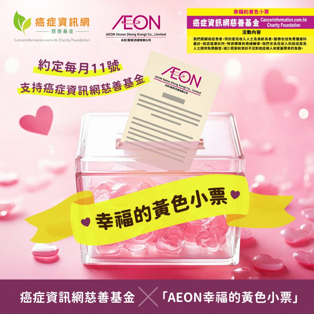
Shopping and Blessings 【AEON x CICF 「幸福的黃色小票」🗳】
You can bless cancer survivors during the New Year! you can support Cancerinformation while purchasing festive items! Next Tuesday (11 February after shopping at AEON store, simply drop your yellow receipts into the designated “Cancerinformation.com.hk Charity Foundation” collection box, or vote online by AEON Mobile APP. AEON will donate goods equivalent to 1% of the total transaction amount to the Cancerinformation.com.hk Charity Foundation (CICF).
What is more, we will set up a promotional booth at the AEON Tai Wo Hau Store to raise public awareness of health. Hope you take this opportunity to learn about health information while you shop. Looking forward to seeing everyone there!
📍 AEON 大窩口店
荃灣荃華街3號悅來坊 B2-B3 號舖
下載AEON Mobile Apps 📱網上購物,亦請在應用程式內進行投票支持
癌症資訊網慈善基金
Cancerinformation.com.hk Charity Foundation