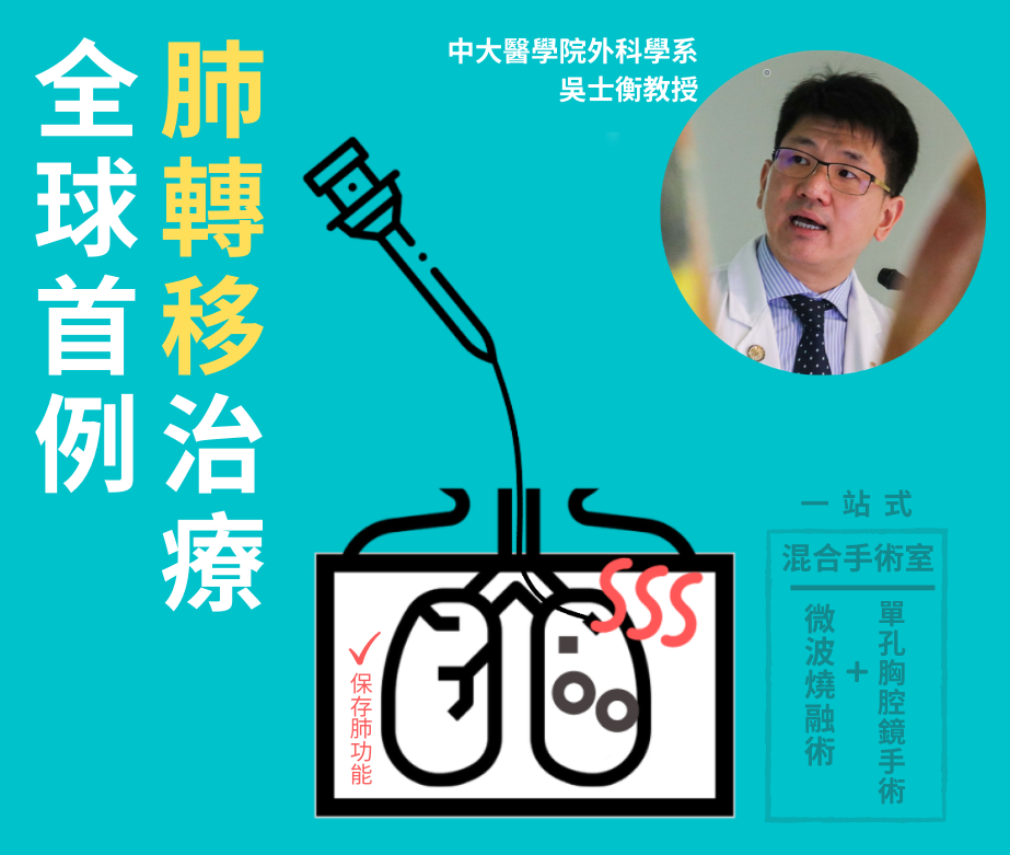
中大醫學院胸腔外科團隊創全球首例肺轉移治療

【全球首例】中大醫學院胸腔外科團隊為肺轉移治療帶來新突破。團隊成功應用混合手術室配合「經氣管微波消融術」及「單孔胸腔鏡肺葉切除術」於同一手術,為一名患有肝癌而出現肺轉移的68歲長者,清除他位於右肺的四個腫瘤組織,成功地保存大部分肺部功能。這是全球首宗利用混合手術室配合多元模式一站式治療肺轉移病人。報告已刊登於國際醫學期刊The Annals of Thoracic Surgery。
中大醫學院外科學系心胸外科組教授吳士衡教授表示:「治療多病灶肺轉一直以來都是莫大挑戰,因為患者有多個腫瘤組織分佈肺部,大小不一,位置及深度都會影響治療選擇。就是次個案而言,以手術切除所有病變組織是最理想的處理方法,但所有腫瘤組織加起來需要切除的肺部面積往往太大,而且操作上亦有困難。因此,我和團隊決定利用混合手術室配合多元模式治療,分別是『經氣管微波消融術』及『單孔胸腔鏡肺葉切除術』,以保存最多肺部組織為目標替病人進行治療。」
團隊首先利用斷層掃描即時確認右肺的四個腫瘤組織位置。當中兩個位於右上肺葉的腫瘤組織,由於體積細小(少於1厘米)而且位置深入,團隊決定以「經氣管微波消融術」破壞病變組織,以無創的方式代替肺葉切除。至於位於右中肺葉近外圍的兩個腫瘤組織較大,則以微創「單孔胸腔鏡肺葉切除術」處理。
吳教授補充:「以往治療有多個腫瘤的肺轉移時,需要分多次手術或治療程序進行,病人需要多次住院及接受麻醉,創傷性較大。混合手術室獨有的實時360度斷層電腦掃描配置讓我們可以在同一手術進行三個程序 — 尖端影像診斷、先進的電磁導航支氣管鏡介入治療和微創手術。這有助為病人度身設計最適切的一站式治療組合,特別適合出現多個及體積少於1厘米的腫瘤組織。此外,在當前新冠肺炎疫情下,一站式的治療可避免患者多次入院,大大減低受感染的機會。」
吳教授及他的團隊為世界著名的混合手術室胸腔外科團隊,在亞太區的「電磁導航支氣管鏡檢查」及「經氣管微波消融術」兩大技術上有著領導性的地位。
報告詳情可參閱:https://bit.ly/2CXsEPv
【World 1st】Thoracic surgical team from CU Medicine achieves breakthrough in the treatment of lung metastasis. The team successfully performed non-invasive bronchoscopic microwave ablation (BMA) and uniportal video-assisted thoracic surgery (uVATS) at a single operation in hybrid operating room for a 68-year-old hepatocellular carcinoma patient with multiple lung metastases, treating four lesions located in different parts of the right lung and preserving most of the lung function. This is world’s first attempt of one-stop hybrid treatment of multiple lung metastases with BMA and uVATS in hybrid operating room. The case report was published in the international medical journal The Annals of Thoracic Surgery.
Professor Calvin NG from CU Medicine’s thoracic surgical team says, “Multifocal lung metastasis is traditionally a challenge to manage. Heterogeneity in size, location and depth of the multiple lesions are decisive factors to treatment options. In this case, ideally all the four metastases should be surgically resected if possible. However, the loss of lung volume precludes our team from considering this as an option and there are other practical difficulties in the resection. Therefore, we decided to implement a multimodal treatment with hybrid theatre facilities, that is to combine BMA and uniportal VATS in one operation, so as to preserve most lung tissues.”
The team confirmed the position of four metastases with cone-beam CT scan, which were on the right middle lobe and right upper lobe. The two lesions on the upper lobe were deep in the anterior segment with size smaller than 1cm and thus BMA was performed to destroy them. For the other two bigger lesions at the periphery of the middle lobe, doctors decided to remove them with uniportal VATS.
“Conventionally, multimodal management for metastases were carried out in separate locations in staged manner that may require patients to have multiple hospital admissions and be anesthetised more than once. The hybrid operating room with cone-beam computed tomography can integrate three procedures into a single operation setting: high-end radiology, state-of-the-art interventional bronchoscopy and advanced minimally invasive surgery. This allows a tailor made one-stop management of lung tumours, particularly for multiple, subcentimeter lesions. Furthermore, avoiding multiple hospital admissions in the current era of COVID-19 could reduce exposure of patients to high risk areas,” Professor Ng added.
Professor Ng and his team is renowned in hybrid operating room thoracic procedures, and pioneers in the use of electromagnetic navigation bronchoscopy and BMA in the Asia Pacific region.
Details of the report: https://bit.ly/2CXsEPv
文章來自: CUHK Medicine


